Oral and Maxillofacial Surgery Textbooks
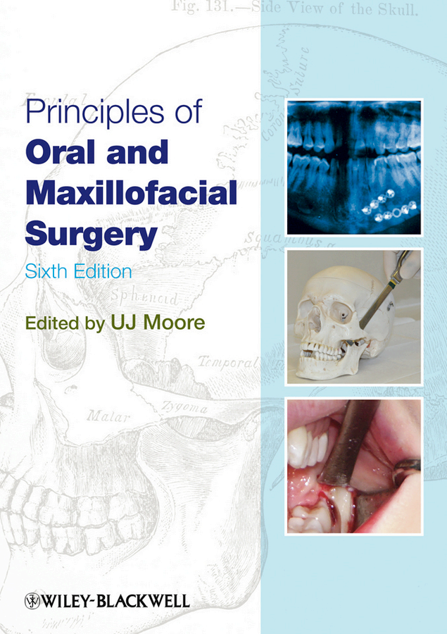
Principles of Oral and Maxillofacial Surgery PDF – UJ Moore
Principles of Oral and Maxillofacial Surgery Textbook Free PDF
Principles of Oral and Maxillofacial Surgery Free PDF
Principles of Oral and Maxillofacial Surgery Free PDF
Principles of Oral and Maxillofacial Surgery by UJ Moore is one of the most respected references in OMFS.
It provides clear explanations of surgical techniques, patient assessment, and management of oral and maxillofacial conditions.
An essential guide for students, residents, and clinicians who wish to strengthen their surgical foundation.
📥 Download the free PDF here:
👉 🔗 Click to Download the Book