
Histopathology of Dental Caries
Histopathology of Dental Caries
👉 Histopathology of Dental Caries MCQs
Dental caries is a dynamic process that begins with demineralization of enamel and may progress into dentin and pulp if untreated.
Histopathologically, caries in enamel shows distinct patterns depending on its location and progression.
1️⃣ Enamel Caries
Caries in enamel typically begins beneath dental plaque deposits. Based on location, it is classified into:
🔹 A. Smooth Surface Caries
- Commonly found on the gingival third of buccal and lingual surfaces, and on proximal surfaces below the contact point.
- Earliest sign: white spot lesion (area of decalcification under plaque). With progression, the area becomes bluish-white.
- Histological changes:
- Loss of interprismatic substance → enamel rods become more prominent.
- Accentuated incremental lines of Retzius.
- Loss of mucopolysaccharides in the organic matrix.
- Lesion pattern: cone-shaped, with apex towards DEJ and base towards the tooth surface.
- Final stage: enamel becomes rough, demineralized, and prism structure disintegrates.
🔹 B. Pit and Fissure Caries
- Pits and fissures are highly susceptible due to their anatomy, which favors bacterial and food entrapment.
- Clinical features: appears brown or black; explorer shows a definite “catch”. Surrounding enamel looks opaque bluish-white.
- Begins under plaque → enamel demineralization.
- Histopathology:
- Caries follows the direction of enamel rods.
- Lesion shape: triangular, with apex at tooth surface and base towards DEJ (opposite to smooth surface caries).
- Thin enamel at fissure base → early dentin involvement is common.
- Leads to undermined enamel; once fractured, cavitation and exposure occur.
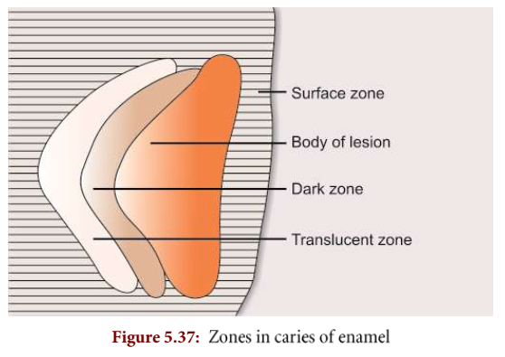
Zones of Enamel Caries (Seen under Polarized Light)
Before complete enamel destruction, four histological zones can be identified:
| Zone | Key Features |
|---|---|
| Translucent Zone | Advancing front of lesion, ~10× more porous than sound enamel, may be absent. |
| Dark Zone | Lies above translucent zone; always present (positive zone); dark under polarized light due to demineralization. |
| Body of the Lesion | Largest area; located between surface zone and dark zone; shows maximum demineralization and porosity. |
| Surface Zone | Least affected; high mineralization & fluoride concentration; <5% porous; radiopacity close to normal enamel. |
📌 Clinical Significance
- Recognizing histological features helps dentists detect caries at an early stage.
- Identifying white spot lesions enables preventive and remineralization therapy rather than operative intervention.
- Understanding lesion progression allows more conservative caries management and preservation of tooth structure.
2️⃣ Dentinal Caries
Although enamel caries is dynamic, it is non-vital (cannot defend itself).
Dentin and pulp are vital tissues, capable of defense. The pulp-dentin complex acts as a structural and functional unit.
When caries reaches the dentinoenamel junction (DEJ):
✓ It spreads laterally (dentin is less resistant than enamel).
✓ Appears brown due to:
● Pigment-producing microorganisms
● Protein breakdown in the presence of sugar
● External stains
Caries in dentin involves:
→ Demineralization of inorganic material
→ Breakdown of collagen fibers
→ Progresses twice as fast as enamel
Early Dentinal Changes (Initial Caries)
✓ Dentinal sclerosis: calcification of tubules → prevents further bacterial penetration
✓ More common in chronic, slow caries
✓ Transparent dentin forms when tubules are fully filled
✓ Intertubular dentin is soft, lumen filled with calcified material
✓ Pioneer bacteria may appear in a few tubules
✓ Fatty degeneration of Tome’s fibers → predisposes to sclerosis
Microbial pattern in dentin:
- Near surface: acid-loving bacteria dominate
- Deeper dentin: proteolytic bacteria dominate (due to protein-rich environment)
Advanced Dentinal Changes
✓ Tubule walls decalcify → tubules merge
✓ Sheath of Neumann may swell irregularly
✓ Tubule diameter increases (packed with bacteria)
✓ Liquefaction foci: ovoid necrotic areas along tubules
✓ Leathery necrotic dentin forms
✓ Clefts perpendicular to tubules → dentin peels in layers
✓ Lesion shape: triangular (apex toward pulp, base toward enamel)
👉 Histopathology of Dental Caries MCQs
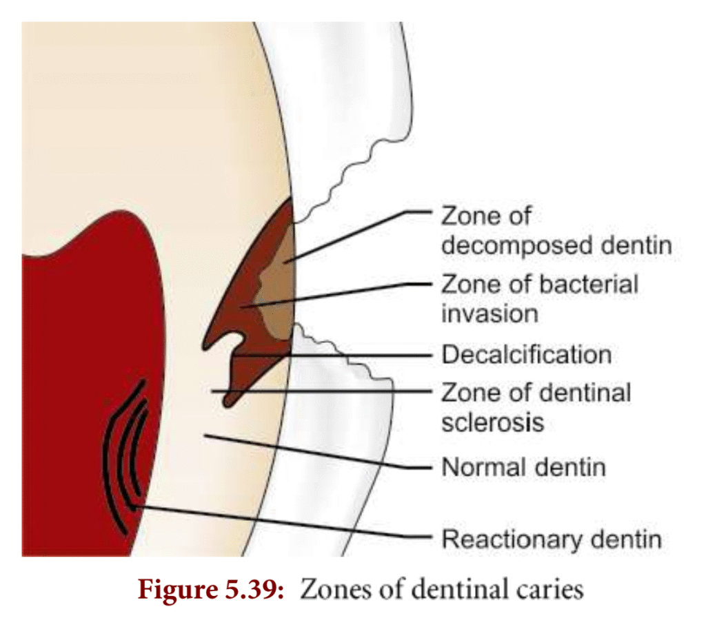
Zones of Dentinal Caries
| Zone | Features | Bacteria | Treatment |
|---|---|---|---|
| Zone 1: Normal dentin | Fatty degeneration of Tome’s fibers; otherwise normal dentin | None | Sensitive; no removal needed |
| Zone 2: Dentinal sclerosis | Intertubular dentin demineralized; tubules filled with Ca salts | None | Can remineralize; no removal needed |
| Zone 3: Decalcified dentin | Softened intertubular dentin | Rare | Monitor; may need removal if progressing |
| Zone 4: Bacterial invasion | Tubules widened/distorted, filled with bacteria | Present | Must remove during tooth prep |
| Zone 5: Decomposed dentin | Outermost dentin, necrotic, filled with bacteria | Present | Must remove completely |