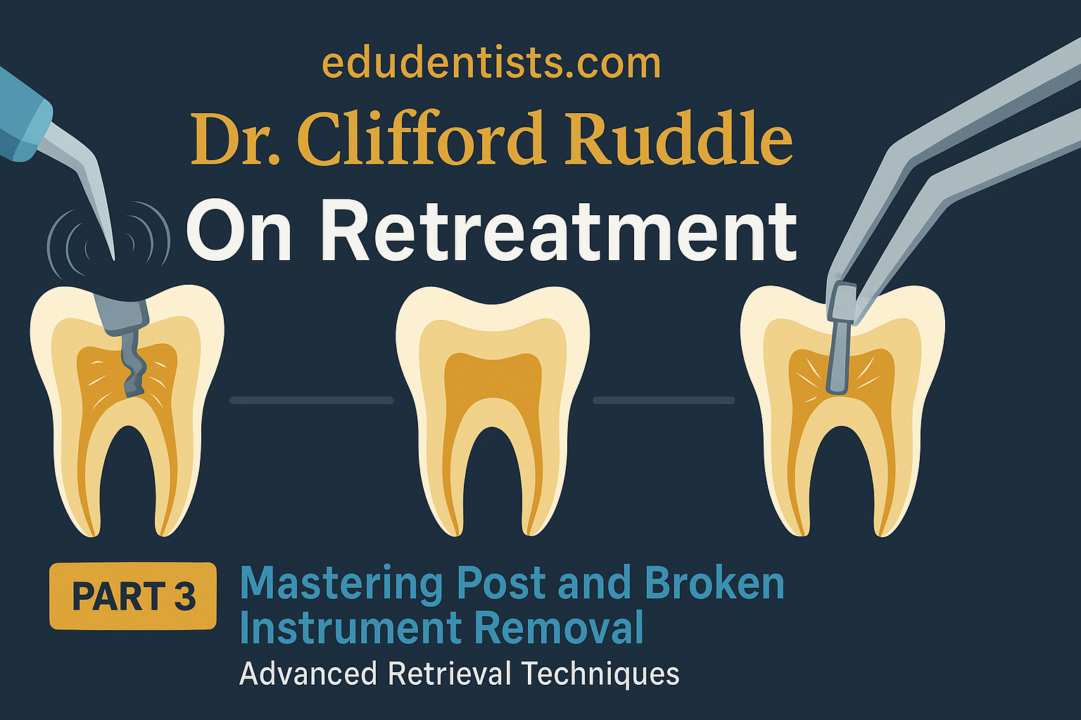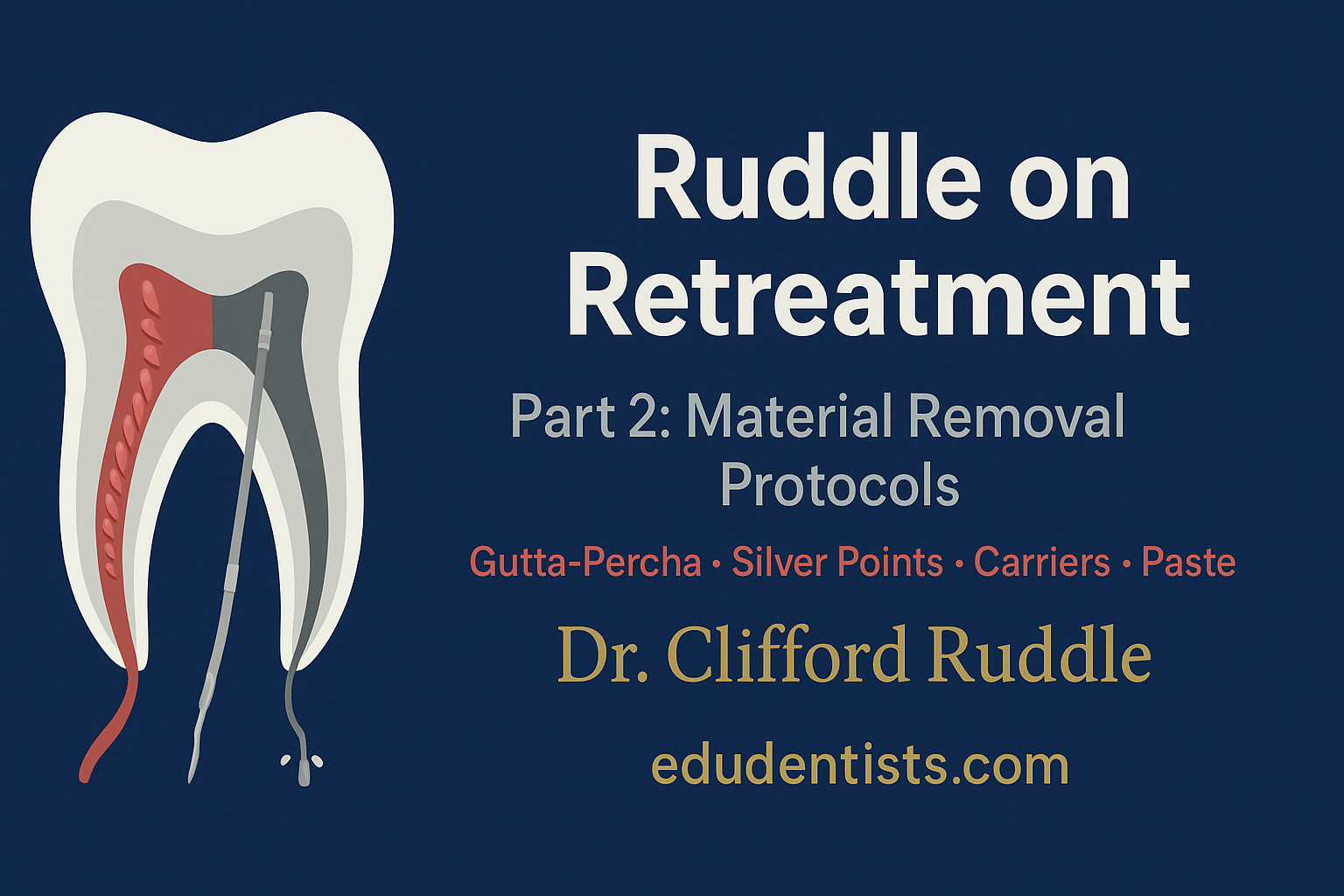
Cleaning and Shaping in Root Canal Treatment
Cleaning and Shaping in Root Canal Treatment

Cleaning and Shaping in Root Canal Treatment
Successful root canal therapy relies on three foundations:
✔ Accurate diagnosis and treatment planning.
✔ Thorough understanding of tooth anatomy.
✔ Effective cleaning, shaping, and sealing of the root canal system.
Principles of Cleaning
Nonsurgical root canal treatment offers a predictable way to retain teeth that might otherwise require extraction.
Key Concepts:
➔ Vital vs. Necrotic Pulp:
- Vital pulp → higher success rates.
- Necrotic pulp with periradicular disease → more challenging due to residual tissue and bacterial by-products.
➔ Limitations of Complete Debridement:
- Complex canal anatomy (lateral canals, fins, cul-de-sacs, isthmuses) makes total cleaning nearly impossible.
- Objective = significant reduction of irritants, not total elimination.
Indicators of Adequate Cleaning:
✦ Smooth, “glassy” canal walls when touched with a small file.
✦ Enlargement 3 file sizes beyond the first binding file.
✦ Irrigant clarity and presence of clean dentinal shavings.
Clinical Note: These measures do not perfectly correlate with cleanliness; the “smooth wall” test is the most reliable.
Principles of Shaping
The goal of shaping is to:
✔ Facilitate cleaning and irrigation.
✔ Create space for obturating materials.
✔ Maintain a continuously tapering funnel from orifice to apex.
Requirements for Shaping:
- Preserve original canal anatomy (avoid ledging, zipping, or transportation).
- Maintain apical foramen in its natural position.
- Develop smooth, tapered walls for obturation.
Shaping Requirements Based on Obturation Method:
| Obturation Method | Shaping Requirement |
|---|---|
| Lateral compaction | Spreaders should penetrate 1–2 mm short of working length |
| Warm vertical compaction | Coronal enlargement should allow pluggers to reach 3–5 mm of working length |
Apical Canal Preparation
One of the most debated aspects of endodontics is where to terminate cleaning and shaping.
Facts about Apical Anatomy:
- Apical constriction (narrowest point, ~0.5 mm coronal to foramen) is irregular and often absent.
- Foramen rarely coincides with anatomic apex.
- Distance between foramen and constriction varies (0.2–3.8 mm).
- Age and resorption alter apical anatomy.
Guidelines:
✦ Terminate preparation 1–3 mm from the radiographic apex.
✦ Success decreases if obturation is >2 mm short or extends beyond the apex.
✦ Extrusion of materials → lower prognosis.
📊 Flowchart: Decision-Making for Apical Termination
        Start
          │
          ▼
   Is pulp necrotic? ──► YES ──► Terminate 0–2 mm short of radiographic apex
          │
          NO
          │
          ▼
  Vital inflamed pulp ──► Terminate 1–3 mm short of radiographic apex
Degree of Apical Enlargement
- Small apical preparation = less risk of transportation but less cleaning.
- Larger apical preparation (≥ #35–40 file) = better irrigation and bacterial reduction.
✦ Balance is key:
✔ Too small → poor disinfection.
✔ Too large → risk of weakening and fracture.
Elimination of Etiology
Mechanical vs. Chemical:
- Mechanical instrumentation alone ≠ sterility.
- Irrigation is essential (volume is more important than concentration).
Common Irrigants:
| Irrigant | Advantages | Disadvantages |
|---|---|---|
| Sodium hypochlorite (NaOCl) | Strong antimicrobial, dissolves organic tissue | Toxic if extruded |
| Chlorhexidine | Antimicrobial, substantivity | Does not dissolve tissue |
Apical Patency
Definition: Passing small files slightly beyond apical foramen.
✔ Advantages: Prevents blockage, maintains working length.
✦ Concerns: May extrude debris, bacteria, or irrigants.
✦ Evidence: No significant bacterial reduction compared to non-patency.
Clinical Insight: Maintaining patency is not biologically essential but may help avoid blockages.
Clinical Pearl: Use selectively — not mandatory in every case.
Pretreatment Evaluation
Before starting, evaluate:
- Root length & curvature (S-shaped or bayonet curvatures are difficult).
- Canal calcification (narrows coronal-apical space).
- Resorption (internal or external).
- Restorations that may obstruct access.
Cleaning and Shaping Techniques
✔ Objectives:
- Develop a tapered funnel.
- Maintain original canal shape.
- Preserve apical foramen in position.
- Avoid over-enlargement.
Instruments & Technology:
| Instrument/Method | Strengths | Limitations |
|---|---|---|
| Stainless steel files | Widely available | Rigid, risk of transportation |
| NiTi rotary files | Flexible, superior shaping | Risk of fracture, costly |
| Ultrasonics | Improves irrigant effectiveness | Limited evidence for smear layer removal |
📊 Flowchart: Instrument Choice in Cleaning & Shaping
       Start
         │
         ▼
 Is canal severely curved? ──► YES ──► Avoid rotary NiTi, use hand files + step-back
         │
         NO
         │
         ▼
 Is canal calcified? ──► YES ──► Consider ultrasonics + gradual hand files
         │
         NO
         │
         ▼
 Standard canal morphology ──► Use rotary NiTi with crown-down technique
Ultrasonics in Endodontics
Uses:
- Cleaning and shaping.
- Post and material removal.
- Root-end preparation.
Mechanism: Acoustic microstreaming → enhances irrigant penetration and debris removal.
Summary
✔ Cleaning and shaping are more important than obturation alone for long-term success.
✔ Apical termination: ideally 1–3 mm short of radiographic apex.
✔ Apical enlargement: #35–40 improves disinfection but must be balanced with tooth strength.
✔ NiTi instruments + irrigants + ultrasonics = modern gold standard, but none achieve sterility alone.
✔ Coronal seal + effective disinfection = ultimate predictors of success.
👉