
Clinical Examination of the Edentulous Patient (Complete Denture) _ Extraoral Examination
Clinical Examination of the Edentulous Patient (Complete Denture) _ Extraoral examination

Clinical Examination of the Edentulous Patient (Complete Denture) _ Part 1 _ Extraoral Examination
The clinical examination is a fundamental step in the dental diagnostic process. It is systematically divided into two main components: the extraoral examination and the intraoral examination.
I. Extraoral Examination
The extraoral examination involves a comprehensive assessment of the patient’s head and neck region for any pathological conditions. Key aspects evaluated include facial color, skin tone, hair color and texture, facial symmetry, and neuromuscular activity. This examination encompasses the following specific areas:
1. Facial Examination
This examination focuses on several critical elements:
- Facial Features: Particular attention is paid to the perioral region, as it significantly influences tooth selection and denture design. The following features are noted:
- Lip length and fullness.
- Apparent lip support.
- The philtrum.
- Nasolabial folds.
- Mentolabial sulcus (labiomental groove).
- Labial commissures and the modiolus.
- Width of the vermillion border (impacts the degree of tooth display).
- Size of the oral opening (also influences tooth display).
- Skin texture (smooth or rough) and color. Rough skin texture may necessitate the selection of more rugged-looking teeth, while wrinkles can indicate a decreased vertical dimension.
These factors collectively guide the selection of the appropriate shade, shape, and arrangement of the artificial teeth.
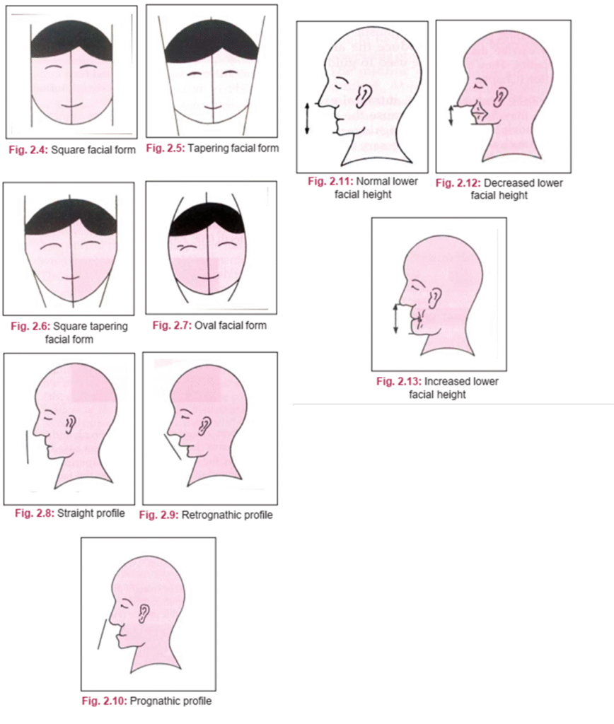
- Facial Form: The outline of the face is classified to aid in teeth selection. Common classifications (by House & Loop, Frush & Fisher, and Williams) include square, tapering, square-tapering, and ovoid forms.
- Facial Profile: The evaluation of the facial profile in the sagittal plane is crucial as it determines jaw relationship and occlusion. Angle’s classification is typically used:
- Class I: Normal or straight profile.
- Class II: Retrognathic profile.
- Class III: Prognathic profile.
- Lower Facial Height: Determining the lower facial height is essential for establishing the correct vertical jaw relation. In edentulous patients wearing existing dentures, the lower facial height is observed during occlusion. A collapsed appearance suggests a loss of vertical dimension (VD), leading to wrinkles around the mouth. Conversely, an excessive VD results in stretched facial tissues.
2. Examination of Muscle Tone and Development
- Muscle Tone: The tone of the masticatory and facial muscles affects denture stability. House’s classification is used:
- Class I: Normal muscle tension and tone, with no degeneration.
- Class II: Normal function but slightly decreased tone.
- Class III: Significantly decreased tone and function, often associated with ill-fitting dentures, decreased VD, reduced biting force, and facial wrinkles.
- Muscle Development: The degree of muscle development correlates with biting force.
- Class I (Heavy)
- Class II (Medium)
- Class III (Light)
3. Complexion
The patient’s eye color, hair color, and skin color provide essential guidance for selecting the shade of artificial teeth. Notably, pale skin may be indicative of anemia.
4. Lip Examination
A detailed lip assessment includes:
- Support: Classified as adequately supported or unsupported.
- Mobility: Classified as normal (Class 1), reduced (Class 2), or paralyzed (Class 3).
- Thickness: Thick lips require less support from the teeth and denture flange, allowing more flexibility in tooth positioning. Thin lips depend heavily on the correct labiolingual placement of teeth for support and fullness.
- Length: A key determinant in anterior teeth selection. Lips are classified as long, medium, or short. Short lips reveal more of the teeth and denture base.
- Health: The lips are inspected for fissures, cracks, or ulcers (e.g., at the corners of the mouth), which may indicate nutritional deficiencies (e.g., Vitamin B), candidiasis, or prolonged overclosure due to decreased VD.
5. Temporomandibular Joint (TMJ) Examination
The TMJ is critical for denture function. The examination assesses the range of motion, presence of pain, condition of the masticatory muscles, and any joint sounds (e.g., clicking) upon opening and closing. Severe TMJ pain can indicate an incorrect vertical dimension.
6. Neuromuscular Examination
This evaluation consists of two parts:
- Speech: Assessed based on the patient’s ability to articulate with existing dentures.
- Type 1 (Normal): Patients with clear speech adapt easily to new dentures.
- Type 2 (Affected): Patients with impaired articulation require special attention during the anterior teeth arrangement and need more time to adapt to new dentures.
- Neuromuscular Coordination: The patient’s overall coordination, gait, and steadiness are observed from the moment they enter the clinic. Abnormalities may indicate underlying neuromuscular disorders (e.g., Parkinson’s disease, hemiplegia, cerebellar diseases) or side effects of psychotropic drugs. Specific abnormal facial movements (e.g., lip smacking, tongue tremors) can compromise denture stability and lead to prosthetic failure. Patients are classified as:
- Class I (Excellent)
- Class II (Fair)
- Class III (Poor)


